
Explore ImmuQuest’s range of high-quality recombinant and traditional monoclonal cancer antibodies for biomarker detection, translational oncology research, immunohistochemistry, flow cytometry and diagnostic assay development.
Diagnostic Applications
Click here to speak with our team about your assay development requirements.
Request a Sample
Click here to discuss purchasing a trial size evaluation sample for diagnostic testing and validation.
Request a Quotation
Click here to request pricing, bulk supply options, or custom antibody requirements.
Key Cancer Biomarker Antibodies
Clinical Tumour Markers
Antibodies targeting established cancer biomarkers, used in tumour detection and disease monitoring. Includes CEA, CA19-9 and other clinically relevant oncology markers.
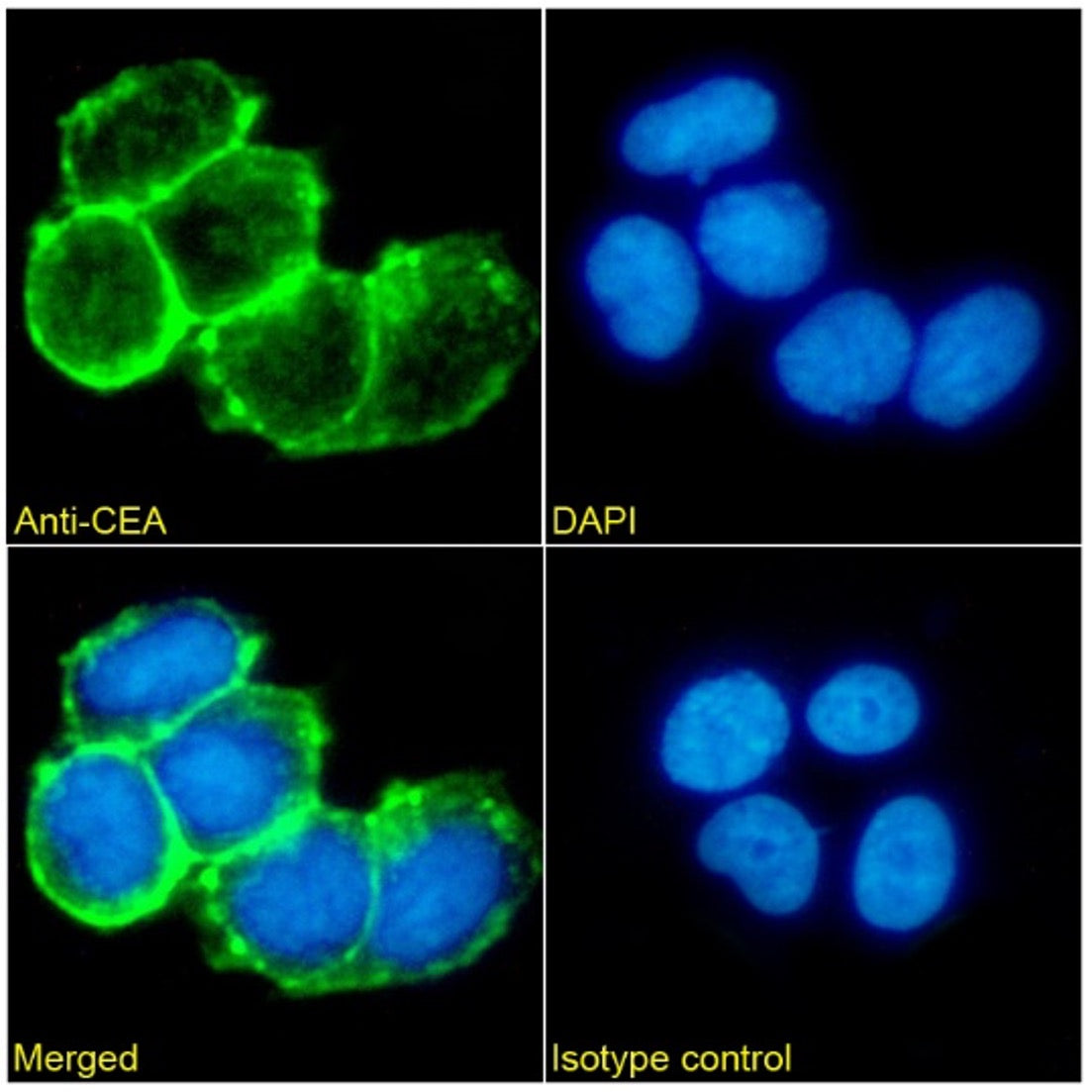
Carcino Embryonic Antigen CEA Monoclonal Antibody [CB30]
CA19-9 Monoclonal Antibody [121SLE]
![Immunohistochemistry (Formalin/PFA-fixed paraffin-embedded sections) - Anti-CA19-9 Antibody [121SLE] Human Colon CA](/cdn/shop/products/1315304723-04146500.jpg?v=1686652367)
ACTH N-Terminal Monoclonal Antibody 6Y8 [57]
![IHC using ACTH N-Terminal Antibody 6Y8 [57] on Human pituitary gland FFPE tissue at 4ug/ml](/cdn/shop/products/1315321437-95537900.jpg?v=1686652394)
DNA Damage Response & Repair Markers
Research antibodies for studying DNA repair, genomic stability and cancer signalling pathways. Includes BRCA2, FANCD2 and NQO1.
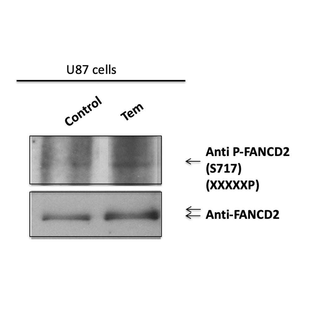
FANCD2 (pSer717) [P-CS-5]
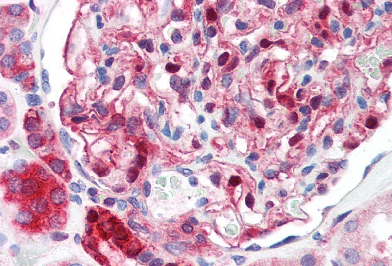
NQO1 Monoclonal Antibody [A180]
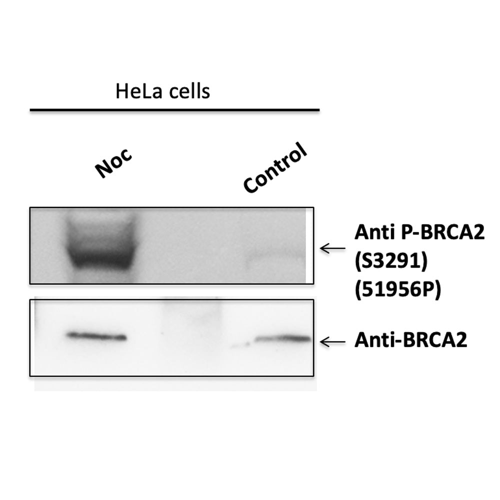
BRCA2 (pSer3291) [P-CS-4]
Tumour Associated Antigens
Antibodies against tumour-associated antigens, epithelial markers and cancer cell surface proteins. Includes Cytokeratins, MUC1, MERTK and related cancer targets.
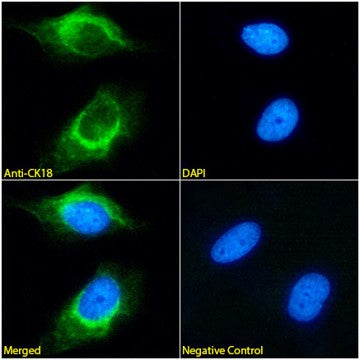
Cytokeratin 18 Monoclonal Antibody [DA-7]
Pan Cytokeratin [C-11], Rabbit IgG1 Kappa, Recombinant Monoclonal Antibody

Mucin-1 Monoclonal Antibody [VU12E1]

Cytokeratin 19 [A53-B/A2], Mouse IgG2a, Lambda Recombinant Monoclonal Antibody
![Cytokeratin 19 [A53-B/A2] Immunofluorescence analysis of paraformaldehyde fixed Hep G2 cells. Primary incubation 1hr (1:100 dilution) followed by Alexa Fluor® 488 secondary antibody (1:1000 dilution), showing cytoplasmic staining. The nuclear stain is DAPI (blue). Isotype control: Ab00178-23 Anti-unknown specificity followed by Alexa Fluor® 488 secondary antibody.](/cdn/shop/files/Cytokeratin_19_A53-B_A2_Immunofluorescence_analysis_of_paraformaldehyde_fixed_Hep_G2_cells._Primary_incubation_1hr_1_100_dilution_followed_by_Alexa_Fluor_488_secondary_antibody_1_1000_dilution_showing_cytoplasmic_staining._The_nuclear_st.png?v=1774449419)
Pan Cytokeratin [C-11], Rabbit IgG1 Kappa, Recombinant Monoclonal Antibody
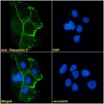
Plakophilin 2 Monoclonal Antibody [8H6]
Leukaemia & Lymphoma Antibodies
Antibodies for haematological malignancy research and immune cell phenotyping. Includes CD and HLA markers for flow cytometry and immunohistochemistry applications.
CD8 [Bu88], Rabbit IgG Kappa, Recombinant Monoclonal Antibody

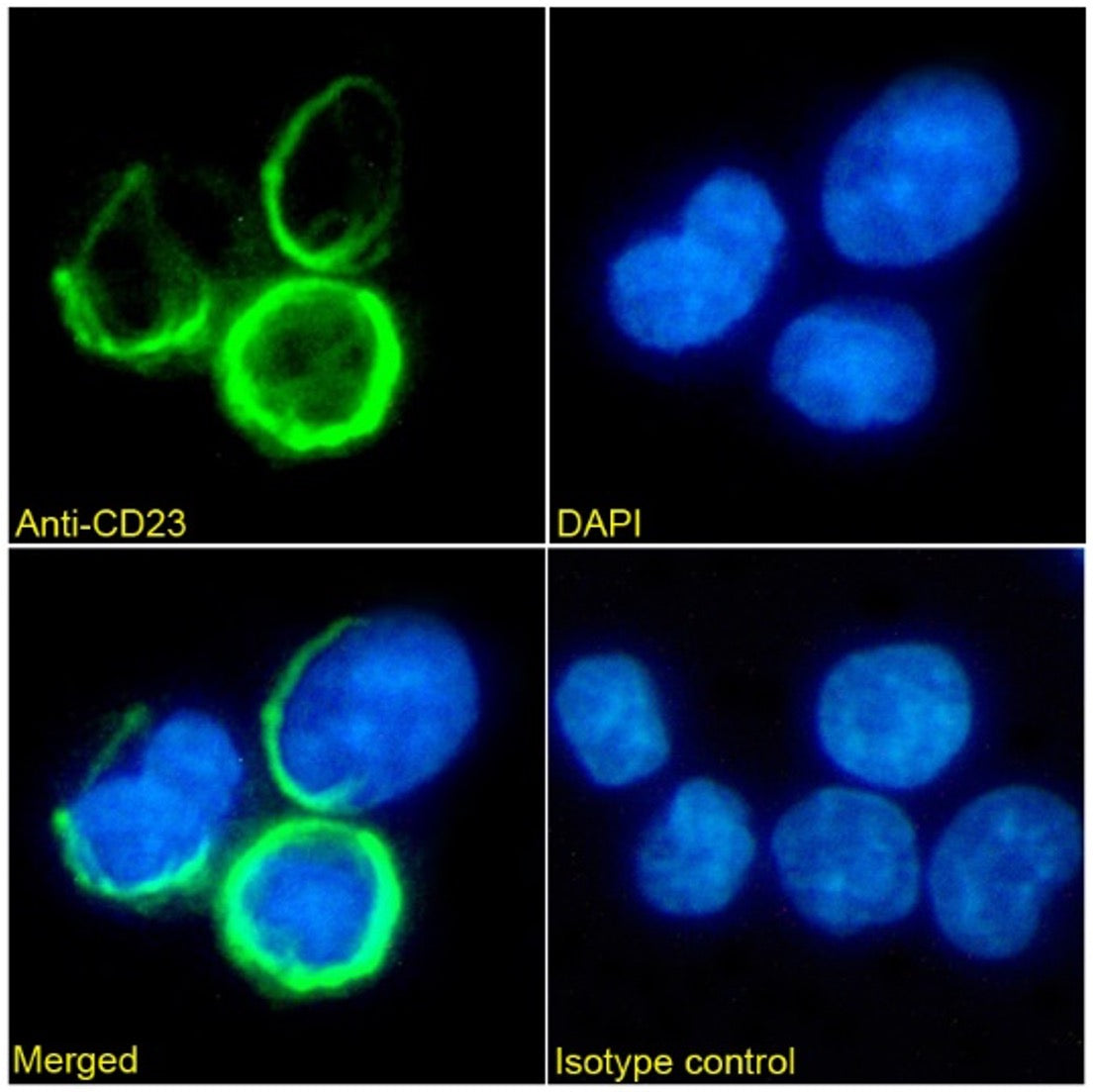
CD23 Monoclonal Antibody [Bu38]
CD33 Monoclonal Antibody [WM53]
![Immunohistochemistry (frozen sections) - Anti-Human CD33 antibody [WM53] normal Human colon tissue](/cdn/shop/products/1362499301-53226900.jpg?v=1606311911)
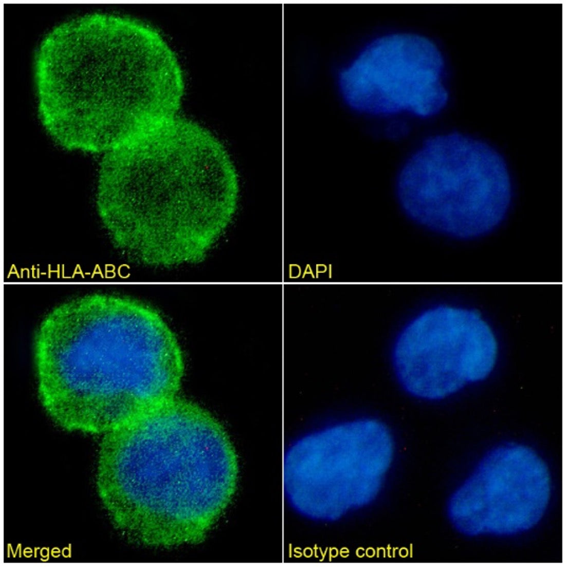
HLA- ABC Monoclonal Antibody [Bu8]
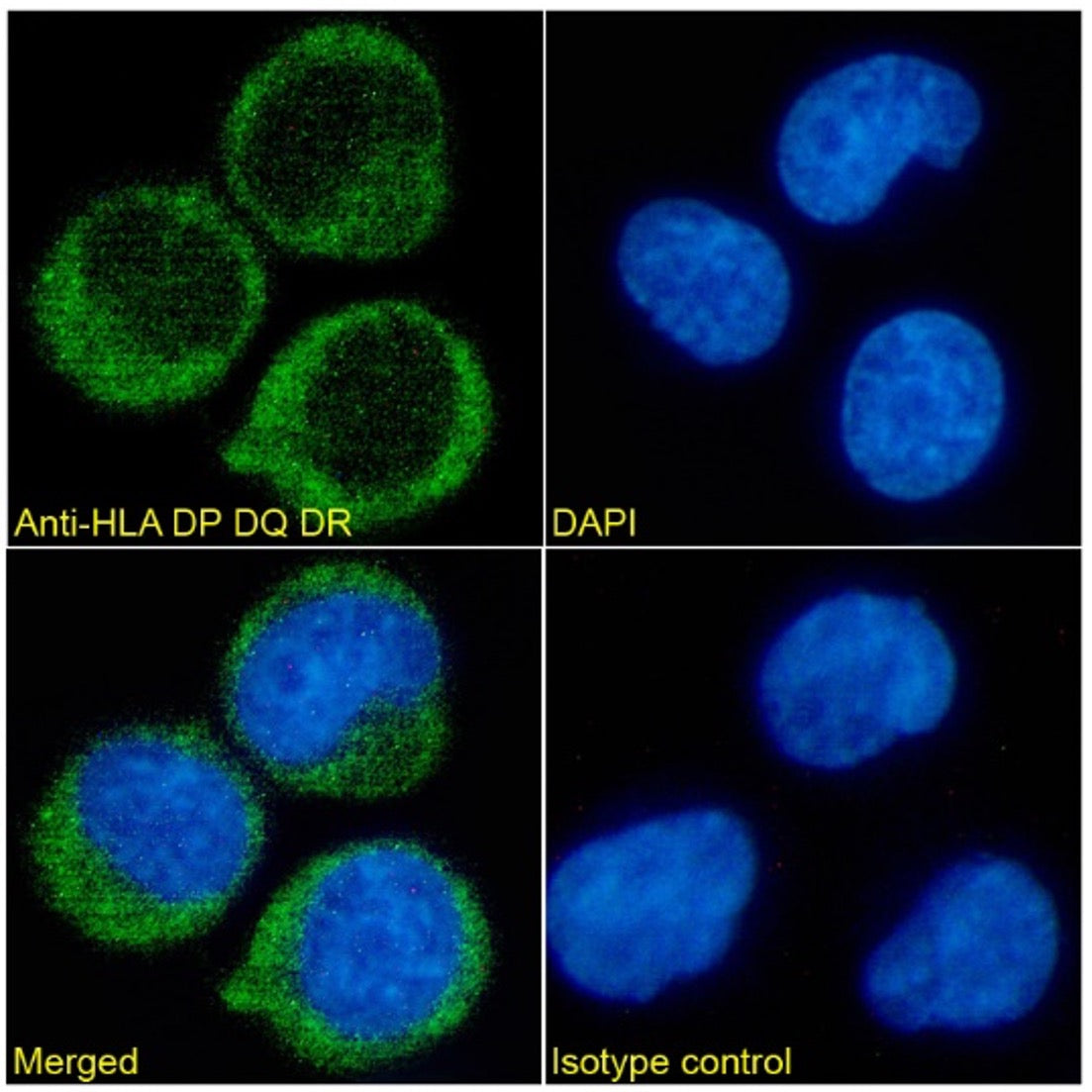
HLA DP DQ DR Monoclonal Antibody [Bu26]
Reproducible Batch Performance
Manufactured for high batch-to-batch consistency, ensuring reliable and reproducible results in diagnostic and research workflows.
Validated Assay
Specificity
Characterised for high specificity and performance across ICC/IF, IHC, Western Blot, Flow Cytometry, and ELISA applications, supporting robust biomarker detection.
Custom Conjugation & Formats
Available with custom conjugation and formats including FITC, HRP, and biotin (BTN), supporting flexible assay design and diagnostic development.
Technical Information & Enquiries
Do you provide technical datasheets for your antibodies?
Yes — detailed datasheets including validation data, applications, and recommended conditions are available upon request.
Can I request antibodies for evaluation in my assay?
Smaller trial-size vials can be purchased for testing in IHC, flow cytometry, ELISA, and related diagnostic workflows.
Are your antibodies suitable for diagnostic and kit development applications?
Many of our antibodies are optimised for use in translational research and diagnostic assay development, including scalable production formats.
Do you offer custom conjugation or specialised formats?
Yes — antibodies are available in formats including FITC, HRP, biotin (BTN), and custom conjugations to support assay design flexibility.
Can I request pricing for bulk or OEM supply?
Yes — we support bulk supply and OEM requirements for diagnostic manufacturers and kit developers.