


![Lamin A+C Monoclonal Antibody [JOL2]](http://immuquest.com/cdn/shop/products/1412848453-24902600_fe29a91f-3e4a-4816-ac2f-41f10d6f59ea_2000x2000.jpg?v=1662642780)


![Lamin A+C Monoclonal Antibody [JOL2]](http://immuquest.com/cdn/shop/products/1412848453-24902600_fe29a91f-3e4a-4816-ac2f-41f10d6f59ea_1000x1000.jpg?v=1662642780)
![Lamin A+C Monoclonal Antibody [JOL2]](http://immuquest.com/cdn/shop/products/1412848453-24902600_fe29a91f-3e4a-4816-ac2f-41f10d6f59ea_{width}x.jpg?v=1662642780)


Current Image Legend:


Lamin A+C Monoclonal Antibody [JOL2]
Catalogue Number: IQ332S
Catalogue Number: IQ608
Catalogue Number: IQ608HRP
Catalogue Number: IQ608FITC
Catalogue Number: IQ332AF
Item in Stock Secure Online Payments
If you have any questions or require further information, please contact us and one of our scientists will be in touch.
A free sample may be available. Please click here to request this.
Datasheet
| Primary Description: | Mouse Anti-Lamin A+C [JOL2] |
| Primary Description: | Lamin A/C Monoclonal Antibody |
| Primary Description: | Mouse Anti-Lamin A+C [JOL2]: Horseradish Peroxidase (HRP) |
| Primary Description: | Lamin A/C Monoclonal Antibody: Fluorescein (FITC) |
| Primary Description: | Lamin A/C Monoclonal Antibody: BSA/Azide Free |
| Target Antigen: | This antibody reacts with both recombinant and native Lamin A and C in humans |
| Target Antigen: | This antibody reacts with both recombinant and native Lamin A and C in humans |
| Target Antigen: | Lamin A+C |
| Target Antigen: | This antibody reacts with both recombinant and native Lamin A and C in humans |
| Target Antigen: | This antibody reacts with both recombinant and native Lamin A and C in humans |
| Catalogue Number: | IQ332S |
| Catalogue Number: | IQ608 |
| Catalogue Number: | IQ608HRP |
| Catalogue Number: | IQ608FITC |
| Catalogue Number: | IQ332AF |
| Quantity: | 0.1ml |
| Quantity: | 0.1mg |
| Quantity: | 50ul |
| Quantity: | 50ul |
| Quantity: | 0.5ml |
| Concentration: | 1mg/ml |
| Clone: | JOL2 |
| Clone: | JOL2 |
| Clone: | JOL2 |
| Clone: | JOL2 |
| Clone: | JOL2 |
| Host: | Mouse |
| Host: | Mouse |
| Host: | Mouse |
| Host: | Mouse |
| Host: | Mouse |
| Isotype: | IgG1 |
| Isotype: | IgG1 |
| Isotype: | IgG1 |
| Isotype: | IgG1 |
| Isotype: | IgG1 |
| Myeloma/Fusion Partners: | Spleen cells from immunised BALB/c mice were fused with cells of the mouse SP2-0/Ag4 myeloma cell line |
| Myeloma/Fusion Partners: | Spleen cells from immunised BALB/c mice were fused with cells of the mouse SP2-0/Ag4 myeloma cell line |
| Myeloma/Fusion Partners: | Spleen cells from immunised BALB/c mice were fused with cells of the mouse SP2-0/Ag4 myeloma cell line |
| Myeloma/Fusion Partners: | Spleen cells from immunised BALB/c mice were fused with cells of the mouse SP2-0/Ag4 myeloma cell line |
| Myeloma/Fusion Partners: | Spleen cells from immunised BALB/c mice were fused with cells of the mouse SP2-0/Ag4 myeloma cell line |
| Immunogen: | Recombinant human lamin A |
| Immunogen: | Recombinant human lamin A |
| Immunogen: | Recombinant human lamin A |
| Immunogen: | Recombinant human lamin A |
| Immunogen: | Recombinant human lamin A |
| Species Reactivity: | Human, African Green Monkey |
| Species Reactivity: | Human, African Green Monkey |
| Species Reactivity: | Human, African Green Monkey |
| Species Reactivity: | Human, African Green Monkey |
| Species Reactivity: | Human, African Green Monkey |
| Purification: | Unpurified |
| Purification: | Protein G |
| Purification: | Unpurified |
| Format: | Tissue culture supernatant containing 0.09% sodium azide |
| Format: | Purified antibody (from supernatant) containing PBS + 0.09% sodium azide |
| Format: | Purified IgG conjugated to Horseradish Peroxidase using InnovaBiosciences Lightning-Link®, supplied in Phosphate buffered saline(PBS) containing 0.01% Thiomersal |
| Format: | Purified antibody conjugated to Fluorescein using Innova Biosciences Lightning-Link®, supplied in Phosphate buffered saline (PBS) containing 0.09% sodium azide |
| Format: | Tissue culture supernatant |
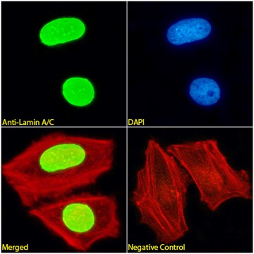
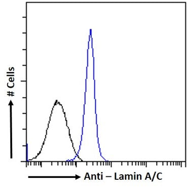
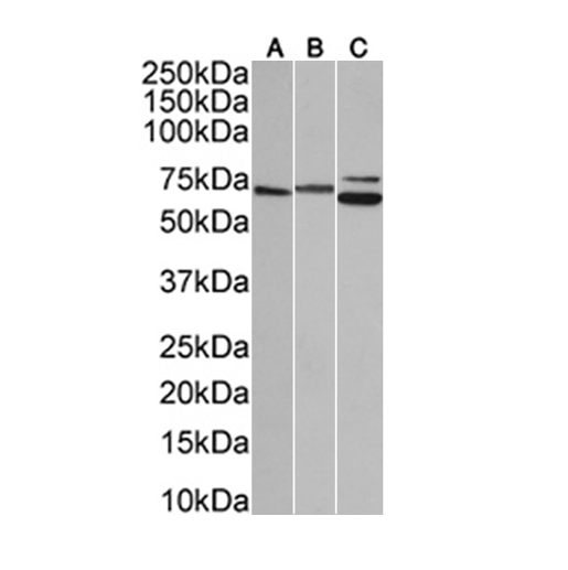
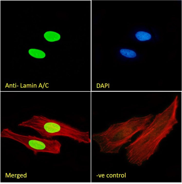
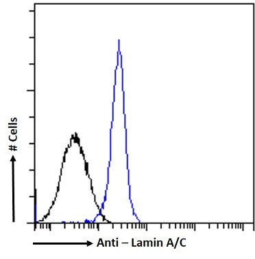
| Applications: | WB, ICC/IF, Flow Cyt |
| Applications: | WB, ICC/IF, Flow Cyt |
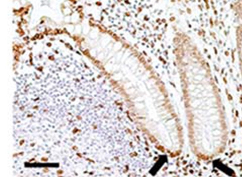
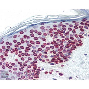
| Applications: | IP, WB, IHC (P) |
| Applications: | WB, ICC/IF, Flow Cyt |
| Applications: | WB, ICC/IF, Flow Cyt |
| Dilutions: | Optimal antibody dilution should be determined by titration, however as a guideline use at: WB 1:1000, IF 1:100, Flow cytometry 1:100 |
| Dilutions: | Optimal antibody dilution should be determined by titration, however as a guideline use at: WB 1:1000, IF 1:100, Flow cytometry 1:100 |
| Dilutions: | Optimal antibody dilution should be determined by titration, however as a guideline try;IHC (P) at 4µg/ml |
| Dilutions: | Optimal antibody dilution should be determined by titration, however as a guideline use at: WB 1:1000, IF 1:100, Flow cytometry 1:1000 |
| Dilutions: | Optimal antibody dilution should be determined by titration, however as a guideline use at: WB 1:1000, IF 1:100, Flow cytometry 1:100 |
| Also Known As: | 70 kDa Lamin antibody;Cardiomyopathy dilated 1A (autosomal dominant) antibody;CDCD1 antibody;CDDCantibody;Charcot Marie Tooth Disease Axonal Type 2B1 antibody;CMD1A antibody;CMT2B1 antibody;EMD2antibody;FPL antibody;FPLD antibody;HGPS antibody;IDC antibody;Lamin A/C antibody;Lamin A/C Isoform 1Precursor antibody;Lamin A/C Isoform 2 antibody;Lamin A/C Isoform 3 antibody;LDP1 antibody;LFPantibody;LGMD1B antibody;Limb girdle muscular dystrophy 1B (autosomal dominant) antibody;LMN 1 antibody;LMN Aantibody;LMN C antibody;LMN1 antibody;LMNA antibody;LMNC antibody;NY REN 32 antigen antibody;PRO1antibody;Progeria 1 (Hutchinson Gilford Type) antibody;Renal carcinoma antigen NY REN 32 antibody;Renalcarcinoma antigen NYREN32 antibody |
| Also Known As: | 70 kDa Lamin antibody;Cardiomyopathy dilated 1A (autosomal dominant) antibody;CDCD1 antibody;CDDCantibody;Charcot Marie Tooth Disease Axonal Type 2B1 antibody;CMD1A antibody;CMT2B1 antibody;EMD2antibody;FPL antibody;FPLD antibody;HGPS antibody;IDC antibody;Lamin A/C antibody;Lamin A/C Isoform 1Precursor antibody;Lamin A/C Isoform 2 antibody;Lamin A/C Isoform 3 antibody;LDP1 antibody;LFPantibody;LGMD1B antibody;Limb girdle muscular dystrophy 1B (autosomal dominant) antibody;LMN 1 antibody;LMN Aantibody;LMN C antibody;LMN1 antibody;LMNA antibody;LMNC antibody;NY REN 32 antigen antibody;PRO1antibody;Progeria 1 (Hutchinson Gilford Type) antibody;Renal carcinoma antigen NY REN 32 antibody;Renalcarcinoma antigen NYREN32 antibody |
| Also Known As: | 70 kDa Lamin antibody;Cardiomyopathy dilated 1A (autosomal dominant) antibody;CDCD1 antibody;CDDCantibody;Charcot Marie Tooth Disease Axonal Type 2B1 antibody;CMD1A antibody;CMT2B1 antibody;EMD2antibody;FPL antibody;FPLD antibody;HGPS antibody;IDC antibody;Lamin A/C antibody;Lamin A/C Isoform 1Precursor antibody;Lamin A/C Isoform 2 antibody;Lamin A/C Isoform 3 antibody;LDP1 antibody;LFPantibody;LGMD1B antibody;Limb girdle muscular dystrophy 1B (autosomal dominant) antibody;LMN 1 antibody;LMN Aantibody;LMN C antibody;LMN1 antibody;LMNA antibody;LMNC antibody;NY REN 32 antigen antibody;PRO1antibody;Progeria 1 (Hutchinson Gilford Type) antibody;Renal carcinoma antigen NY REN 32 antibody;Renalcarcinoma antigen NYREN32 antibody |
| Also Known As: | 70 kDa Lamin antibody;Cardiomyopathy dilated 1A (autosomal dominant) antibody;CDCD1 antibody;CDDCantibody;Charcot Marie Tooth Disease Axonal Type 2B1 antibody;CMD1A antibody;CMT2B1 antibody;EMD2antibody;FPL antibody;FPLD antibody;HGPS antibody;IDC antibody;Lamin A/C antibody;Lamin A/C Isoform 1Precursor antibody;Lamin A/C Isoform 2 antibody;Lamin A/C Isoform 3 antibody;LDP1 antibody;LFPantibody;LGMD1B antibody;Limb girdle muscular dystrophy 1B (autosomal dominant) antibody;LMN 1 antibody;LMN Aantibody;LMN C antibody;LMN1 antibody;LMNA antibody;LMNC antibody;NY REN 32 antigen antibody;PRO1antibody;Progeria 1 (Hutchinson Gilford Type) antibody;Renal carcinoma antigen NY REN 32 antibody;Renalcarcinoma antigen NYREN32 antibody |
| Pubmed ID(s): | 31048846O, 30422822, 31847370, 30126195, 29405587, 28668644, 29156644, 28985428, 28031327, 28179995, 27378237, 27031510, 27926867, 26124276, 26447202, 24452336, 24659496, 24838313, 24950247, 24732130, 23975040, 22871573, 22253444, 21444690, 21610090, 21852285, 21392397, 22040608, 19623164, 11207047, 11683386 |
| Pubmed ID(s): | 30650545, 31048846, 30422822, 31847370, 30126195, 29405587, 28668644, 29156644, 28985428, 28031327, 28179995, 27378237, 27031510, 27926867, 26124276, 26447202, 24452336, 24659496, 24838313, 24950247, 24732130, 23975040, 22871573, 22253444, 21610090, 21852285, 21392397, 22040608, 19623164, 11207047 |
| Pubmed ID(s): | 11683386 |
| Pubmed ID(s): | 30650545, 31048846, 30422822, 31847370, 30126195, 29405587, 28668644, 29156644, 28985428, 28031327, 28179995, 27378237, 27031510, 27926867, 26124276, 26447202, 24452336, 24659496, 24838313, 24950247, 24732130, 23975040, 22871573, 22253444, 21610090, 21852285, 21392397, 22040608, 19623164, 11207047 |
| Pubmed ID(s): | 30650545, 31048846, 30422822, 31847370, 30126195, 29405587, 28668644, 29156644, 28985428, 28031327, 28179995, 27378237, 27031510, 27926867, 26124276, 26447202, 24452336, 24659496, 24838313, 24950247, 24732130, 23975040, 22871573, 22253444, 21610090, 21852285, 21392397, 22040608, 19623164, 11207047 |
| Entrez Gene ID(s): | 103223838, 4000, 25437 |
| Entrez Gene ID(s): | 103223838, 4000 |
| Entrez Gene ID(s): | 103223838, 4000, 24672 |
| Entrez Gene ID(s): | 103223838, 4000 |
| Entrez Gene ID(s): | 103223838, 4000 |
| SwissProt ID(s): | p02545, Q63190 |
| SwissProt ID(s): | p02545 |
| SwissProt ID(s): | p02545, P63331 |
| SwissProt ID(s): | p02545 |
| SwissProt ID(s): | p02545 |
| Omim ID(s): | 150330 |
| Omim ID(s): | 150330 |
| Omim ID(s): | 150330 |
| Omim ID(s): | 150330 |
| Omim ID(s): | 150330 |
Citations
| Citation Count: | 38 |
| Citations: | Mohammadian Gol T et al. Depletion of Akt1 and Akt2 Impairs the Repair of Radiation-Induced DNA Double Strand Breaks via Homologous Recombination. Int J Mol Sci 20:N/A (2019). Blenski M & Kehlenbach RH Targeting of LRRC59 to the Endoplasmic Reticulum and the Inner Nuclear Membrane. Int J Mol Sci 20:N/A (2019).PubMed: 30650545Bensalah M et al. Combined methods to evaluate human cells in muscle xenografts. PLoS One 14:e0211522 (2019). smanagic-Myers S et al. Endothelial progerin expression causes cardiovascular pathology through an impaired mechanoresponse. J Clin Invest 129:531-545 (2019). Borroni AP et al. Smurf2 regulates stability and the autophagic-lysosomal turnover of lamin A and its disease-associated form progerin. Aging Cell 17:N/A (2018). WB ; Human . Tiwari A et al. Stress-Induced Phosphorylation of Nuclear YB-1 Depends on Nuclear Trafficking of p90 Ribosomal S6 Kinase. Int J Mol Sci 19:N/A (2018). Duclos CM et al. Caspase-mediated proteolysis of the sorting nexin 2 disrupts retromer assembly and potentiates Met/hepatocyte growth factor receptor signaling. Cell Death Discov 3:16100 (2017). Human . Sierra Potchanant EA et al. INPP5E Preserves Genomic Stability through Regulation of Mitosis. Mol Cell Biol 37:N/A (2017). Qi L et al. An RNA editing/dsRNA binding-independent gene regulatory mechanism of ADARs and its clinical implication in cancer. Nucleic Acids Res 45:10436-10451 (2017). Mueck K et al. Akt1 Stimulates Homologous Recombination Repair of DNA Double-Strand Breaks in a Rad51-Dependent Manner. Int J Mol Sci 18:N/A (2017). Bergqvist C et al. An inner nuclear membrane protein induces rapid differentiation of human induced pluripotent stem cells. Stem Cell Res 23:33-38 (2017). ICC/IF ; Human . Chen JC et al. Morpholino-mediated Knockdown of DUX4 Toward Facioscapulohumeral Muscular Dystrophy Therapeutics. Mol Ther 24:1405-11 (2016). Fahrenkrog B et al. Expression of Leukemia-Associated Nup98 Fusion Proteins Generates an Aberrant Nuclear Envelope Phenotype. PLoS One 11:e0152321 (2016). Comaills V et al. Genomic Instability Is Induced by Persistent Proliferation of Cells Undergoing Epithelial-to-Mesenchymal Transition. Cell Rep 17:2632-2647 (2016). Garcia-Belinchón M et al. An Early and Robust Activation of Caspases Heads Cells for a Regulated Form of Necrotic-like Cell Death. J Biol Chem 290:20841-55 (2015). Human . Mitchell MJ et al. Lamin A/C deficiency reduces circulating tumor cell resistance to fluid shear stress. Am J Physiol Cell Physiol 309:C736-46 (2015). ICC/IF ; Human . Zhang Y et al. Human skeletal muscle xenograft as a new preclinical model for muscle disorders. Hum Mol Genet N/A:N/A (2014). Human . Ferreboeuf M et al. Nuclear protein spreading: implication for pathophysiology of neuromuscular diseases. Hum Mol Genet N/A:N/A (2014). Sánchez-Osuna M et al. Caspase-activated DNase is necessary and sufficient for oligonucleosomal DNA breakdown, but not for chromatin disassembly during caspase-dependent apoptosis of LN-18 glioblastoma cells. J Biol Chem 289:18752-69 (2014). Human . Jafferali MH et al. MCLIP, an effective method to detect interactions of transmembrane proteins of the nuclear envelope in live cells. Biochim Biophys Acta 1838:2399-403 (2014). WB ; Human . Camps J et al. Loss of lamin B1 results in prolongation of S phase and decondensation of chromosome territories. FASEB J 28:3423-34 (2014). Vallese D et al. The Rag2?Il2rb?Dmd? mouse: a novel dystrophic and immunodeficient model to assess innovating therapeutic strategies for muscular dystrophies. Mol Ther 21:1950-7 (2013). Iglesias-Guimarais V et al. Apoptotic DNA Degradation into Oligonucleosomal Fragments, but Not Apoptotic Nuclear Morphology, Relies on a Cytosolic Pool of DFF40/CAD Endonuclease. J Biol Chem 287:7766-79 (2012). WB ; Human Krom YD et al. Generation of isogenic D4Z4 contracted and noncontracted immortal muscle cell clones from a mosaic patient: a cellular model for FSHD. Am J Pathol 181:1387-401 (2012). Iglesias-Guimarais V et al. Apoptotic DNA Degradation into Oligonucleosomal Fragments, but Not Apoptotic Nuclear Morphology, Relies on a Cytosolic Pool of DFF40/CAD Endonuclease. J Biol Chem 287:7766-79 (2012). WB ; Human . Mamchaoui K et al. Immortalized pathological human myoblasts: towards a universal tool for the study of neuromuscular disorders. Skelet Muscle 1:34 (2011). ICC/IF ; Human Toulany M et al. Impact of oncogenic K-RAS on YB-1 phosphorylation induced by ionizing radiation. Breast Cancer Res 13:R28 (2011). WB ; Human Harten IA et al. Age-Dependent Loss of MMP-3 in Hutchinson-Gilford Progeria Syndrome. J Gerontol A Biol Sci Med Sci : (2011) Gudise S et al. Samp1 is functionally associated with the LINC complex and A-type lamina networks. J Cell Sci 124:2077-85 (2011) Harrigan JA et al. Replication stress induces 53BP1-containing OPT domains in G1 cells. J Cell Biol 193:97-108 (2011). ICC/IF ; Human Harrigan JA et al. Replication stress induces 53BP1-containing OPT domains in G1 cells. J Cell Biol 193:97-108 (2011). ICC/IF ; Human . Gudise S et al. Samp1 is functionally associated with the LINC complex and A-type lamina networks. J Cell Sci 124:2077-85 (2011). Harten IA et al. Age-Dependent Loss of MMP-3 in Hutchinson-Gilford Progeria Syndrome. J Gerontol A Biol Sci Med Sci : (2011). Toulany M et al. Impact of oncogenic K-RAS on YB-1 phosphorylation induced by ionizing radiation. Breast Cancer Res 13:R28 (2011). WB ; Human . Mamchaoui K et al. Immortalized pathological human myoblasts: towards a universal tool for the study of neuromuscular disorders. Skelet Muscle 1:34 (2011). ICC/IF ; Human . Negroni E et al. In vivo myogenic potential of human CD133+ muscle-derived stem cells: a quantitative study. Mol Ther 17:1771-8 (2009). ICC/IF ; Human Negroni E et al. In vivo myogenic potential of human CD133+ muscle-derived stem cells: a quantitative study. Mol Ther 17:1771-8 (2009). ICC/IF ; Human . Both emerin and lamin C depend on lamin A for localization at the nuclear envelope. Vaughan A ( J Cell Sci,Jul 2001) Venables RS et al. Expression of individual lamins in basal cell carcinomas of the skin. Br J Cancer 84:512-9 (2001). |
Protocols
Immunofluorescence protocol - Formaldehyde fixation
- Collect cells from T.c.unit and remove media from petri dish using suction.
- Wash with 1x PBS and remove.
- Incubate cells in pre-warm (37°C) Para-Formaldehyde for 12 minutes at room temperature on an orbital shaker.
- Remove PFA and incubate in 0.5% Triton X-IOO in 1x PBS for 5 minutes at room temperature.
- Prepare blocking reagent, this is also the antibody diluent.
- Wash cells 2x with 1x PBS at room temperature, for 4 minutes/wash on an orbital shaker.
- Block with 1 % NCS and 1x PBS for 30 minutes at room temperature.
- Prepare primary antibodies (50?l/coverslip) and moist staining chambers.
- Wash cells 2x with lx PBS at room temperature and air dry briefly.
- Incubate with primary antibody for 1 hr at room temperature in the dark in staining chambers. During this time prepare the secondary antibody.
- Wash cells 5x with 1x PBS (5 beaker changes/5 counts in each beaker)
- Incubate with secondary antibody for 1 hour at room temperature in the dark in staining chambers.
- Wash cells 5x with 1x PBS.
- Mount in Dapi.
Solutions (prepare fresh the same day of staining).
- * 1x Phosphate buffered saline.
- * Blocking reagent: 1% NCS in 1x PBS (use fresh l0x PBS).
- * Fixation solution: 3.5% Para formaldehyde.
1.75g PFA in 20 ml d.H20 plus 5 drops 1M NaOH. Stir on a hot plate at 50-60°C until dissolved. Add 4 drops IN HCI and check pH indicator strip. PH should be 7.4. Complete volume with d.H20 to 25ml and add 25ml 2xPBS. Check pH before adding to cover slips.
Immunofluorescence protocol - Methanol/acetone fixation
- Collect cells from T.C.unit and remove media from petri dish using suction.
- Wash with 1x PBS and remove.
- Fix cells with cold methanol: acetone 1: 1 for 10 minutes on ice.
- Prepare blocking reagent, this is also the diluent for the antibodies.
- Remove fixative and wash cells 3x with Ix PBS at RT, for 4 minutes/wash on orbital shaker.
- Block with 1% NCS and Ix PBS for 30 minutes at RT.
- Prepare primary antibodies (50?l/coverslip) and moist staining chambers.
- Wash cells 2x with 1 x PBS at RT and air dry for approximately 7 minutes.
- Incubate with primary antibody for 1 hr at RT in the dark in staining chambers. During this time prepare secondary antibody.
- Wash cells 5x with 1x PBS (5 beaker changes/5 counts in each beaker)
- Incubate with secondary antibody for 1 hr at R T in the dark in staining chambers.
- Wash cells 5x with 1x PBS.
- Mount in Dapi.
Solutions (prepare fresh the same day of staining)
- * 1x Phosphate buffered saline.
- * Blocking reagent: 1% NCS in 1x PBS (use fresh 10x PBS).
- * Fixation solution: methanol:acetone 1: 1 ice cold.
Western Blotting Protocol
- Transfer gel to PDVF or nitrocellulose membrane
- Place membrane in plastic tray in blocking buffer for one hour with agitation
- Rinse in wash buffer
- Incubate in wash buffer plus primary antibody for one hour
- Wash 6 X 5 minutes with wash buffer
- Incubate in wash buffer plus secondary antibody for one hour
- Wash 6X 5 minutes with wash buffer
- Detect (e.g. ECL, Amersham according to manufacturers instructions)
Wash buffer
PBS + 0.1% Tween 20
Blocking buffer
Wash buffer + 5% dried milk powder
The concentration of antibodies used depends on each antibody, the amount of antigen and the detection method used. Generally, dilution is in the range of a few hundred times dilution to a few thousand times dilution, but usually has to be determined empirically.
FAQ's
How do I place an order?
When placing an order we require a purchase order number, plus name and contact details of the purchaser, and the person who will be using the product. We will add VAT for every order received from an EU country, unless you are VAT exempt. For such VAT exempt customers we will need to receive a copy of the exemption certificate with the order.
Orders can be placed either by our website, via email or by mail. Please see our contact us page for more details
All orders are subject to availability. Prices of products do not include shipping, VAT or import duties, where these are applicable.
Price and other information provided are subject to change without notice. While every effort is made to keep information provided Up to date, ImmuQuest will not be liable if errors should occur in such information.
Formal acceptance of an order will take place when the goods are dispatched. If prices should be changed between the time of receipt of an order and dispatch, ImmuQuest will contact you in advance.
Ordering via the website
To order via the website you will need to find the product(s) you are looking for by either using our search, browsing our categories or using our full antibody catalogue.
Once you have found the antibodies you are looking for you can view more detailed information about a specific antibody by clicking the product or the 'View' button. Once you are sure it is the antibody you are looking for you can add it to your order by clicking the 'Add To Order' button.
This places the antibody in your order basket, you can add as many antibodies as you wish. Once you have selected all of the antibodies you wish to order then click the checkout button. If you are not logged in you will be asked to login or to register, if you are logged in you will be taken to the checkout page.
On the checkout page you are able to double check your order and enter a purchase order number if you have one.
Once you are happy your order is correct you can purchase the order with debit/credit card details and your order will be submitted. Once we have received and checked your order we will contact you to arrange payment and delivery.
How long does delivery take?
Delivery usually takes no more than one week from reciept of an order.
Do you guarantee your antibodies?
Yes. Should any product not perform as described in the product literature, it will be replaced or a full refund will be given. The customer must notify ImmuQuest within 30 days after the goods have been received to request a replacement, forwarding complete test data as requested by ImmuQuest.
If items are ordered incorrectly by the customer, ImmuQuest will consider taking them back as long as they have been stored correctly and have not been opened or tampered with. Such orders may be subject to a 15% restocking charge on the items plus any shipping costs.
Requests for returns must have prior authorization from ImmuQuest, and must be made within 7 days of receipt of the items. Items must be returned in the same or equivalent packaging as originally dispatched, and by an equivalent method of delivery.
Do you charge VAT?
If you are in the EU and you are no exempt from paying VAT then we must charge VAT (charged at the prevailing UK rate).
Customers that are exempt need to fax an exemption form showing their vat exemption number.
What if I don't have a purchase order number?
You can still place an order without providing a purchase order number but we must recieve payment before your order is despatched.
Non-website orders
Non-website orders can be placed directly with Immuquest at sales@immuquest.com or click on the "contact us" tab for address details
Delivery
Offline Orders
For offline orders please call +44 (0) 1642 713533 or click here to email us.
Delivery & returns
We use federal Express for UK & international shipments, andfor standard shipments the cost is £30 UK & £55 international. If the customer uses their account, we charge a £20 handling fee (UK & international)
We are strongly committed to providing the best quality products, and the best service possible.
Should any product not perform as described in the product literature, it will be replaced or a full refund will be given. The customer must notify ImmuQuest within 30 days after the goods have been received to request a replacement, forwarding complete test data as requested by ImmuQuest.
If items are ordered incorrectly by the customer, ImmuQuest will consider taking them back as long as they have been stored correctly and have not been opened or tampered with. Such orders may be subject to a 15% restocking charge on the items plus any shipping costs.
Requests for returns must have prior authorization from ImmuQuest, and must be made within 7 days of receipt of the items. Items must be returned in the same or equivalent packaging as originally dispatched, and by an equivalent method of delivery.
Product Guarantee
We are strongly committed to providing the best quality products, and the best service possible. In order to do this we depend on your feedback.
Should any product not perform as described in the product literature, it will be replaced or a full refund will be given. The customer must notify ImmuQuest within 30 days after the goods have been received to request a replacement, forwarding complete test data as requested by ImmuQuest.
If items are ordered incorrectly by the customer, ImmuQuest will consider taking them back as long as they have been stored correctly and have not been opened or tampered with. Such orders may be subject to a 15% restocking charge on the items plus any shipping costs.
Requests for returns must have prior authorization from ImmuQuest, and must be made within 7 days of receipt of the items. Items must be returned in the same or equivalent packaging as originally dispatched, and by an equivalent method of delivery.
If you have any questions please contact us using the following details:
ImmuQuest Ltd
Springboard
Stokesley Business Park
24 Ellerbeck Way
Stokesley
TS9 5JZ
UK
Tel: 01642 713533
Fax: 01642 713988
International Tel: +44 1642 713533
International Fax: +44 1642 713988
When placing an order ImmuQuest require a purchase order number, plus name and contact details of the purchaser, and the person who will be using the product. ImmuQuest will also need a VAT number for customers in the European Union.UK customers that are VAT exempt need to fax an exemption certificate.
Orders can be placed either by our website, via email or by mail. Please see our contact us page for more details. All orders are subject to availability. Prices of products do not include shipping, VAT or import duties where these are applicable.
Price and other information provided are subject to change without notice. While every effort is made to keep information provided Up to date, ImmuQuest will not be liable if errors should occur in such information. Formal acceptance of an order will take place when the goods are dispatched. If prices should be changed between the time of receipt of an order and dispatch, ImmuQuest will contact you in advance.


